The aesthetic surgeon must possess a thorough understanding of the brow and forehead anatomy, as well as a comprehension of the dynamic interrelationship between the forehead, brow, eyelid, and midface. The surgeon must also be familiar with the various methods available to effect changes in these tissues, as no single technique enjoys universal application (Table 6-1).
This chapter will provide the reader a thorough review of the anatomy of the brow and forehead, review the most common techniques for rejuvenation, and provide an algorithm from which to apply them.
Jeffrey E. Janis, Jason K. Potter and Rod J. Rohrich
Aesthetics
The absolute dimension of the forehead as measured from glabella to trichion, varies from patient to patient. The general characteristics of a pleasing brow have been well described.1,2 The brow and forehead create the upper one third of the face in the aesthetically proportioned face. The anterior hairline is typically 5—6 cm above the brow. The eyebrow forms a subtle arc that peaks at the junction of the middle and lateral thirds, which should correspond to a point above the lateral limbus. This arc is flatter in males. In females, the brow should be 3-5 mm above the superior orbital rim. In males, it should lie at the level of the orbital rim. Medially, the brow should begin at a line drawn perpendicular to the lateral aspect of the ala and passing through the medial canthus. The lateral brow is positioned slightly higher than the medial brow and should end at a point on a line drawn obliquely through the ala and lateral canthus (Fig. 6-1).
Anatomy
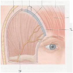
Landmarks for brow position are based upon the underlying bony anatomy. The superior orbital rim is easily palpable and serves as a fixed position for which to assess brow ptosis. Laterally, the temporal ridge delineates the border of the forehead from the temporal fossa (Fig. 6-2). Knize has identified the consistent relationship of several soft tissue structures to the temporal ridge. In this location the soft tissue layers of the forehead and scalp fuse with the periosteum at the zone of fixation (Figs 6-3 & 6-4).

Figure 6-1 Spatial relationships of the ideal eyebrow. Modified from Westmore; reproduced with permission from Ellenbogen R: Transcoronal eyebrow lift with concomitant upper blepharoplasty. Plast Reconstr Surg 1983; 71:490.

Figure 6-2 The forehead and temple subunits. Note that the superior temporal line separates the central forehead from the lateral temporal regions. A, central; B, lateral (temporal); C, eyebrow.

A very important and easily overlooked anatomic variable to consider is calvarial thickness, as many of the techniques of brow lifting involve placement of bony fixation to suspend the newly elevated brow. Calvarial thickness may be as thin as 1-2 mm in the temporal region and along the course of the middle meningeal artery.
The soft tissue anatomy of the forehead, similar to other regions of the face and neck, is arranged in multiple often very subtle layers (Fig. 6-5). The upper forehead is arranged similarly to the scalp with well-defined layers consisting of skin, subcutaneous tissue, galea aponeurosis, loose areolar tissue, and periosteum. At the origin of the frontalis the galea aponeurosis splits into a superficial and deep plane to encase this musculature. The deep plane splits again in the midforehead region to surround the galeal fat pad, and caudal to the fat pad splits again to form the glide plane space of the brow. The periosteum, subgaleal space, and deep galeal plane are discrete layers except in the lower forehead where these layers fuse and are firmly affixed to the frontal bone. Similarly, the periosteum is relatively loosely attached to the frontal bone over the upper and midforehead, but is firmly attached across the lower forehead.
Movement of the brow is produced through the action of brow elevators and depressors, and is enhanced by the presence of the galeal fat pad, glide plane space, and subgaleal space. The major elevator of the brow is the paired frontalis muscle. The frontalis originates from the galeal aponeurosis and inserts into the dermis of the lower forehead. At the level of its insertion, the frontalis interdigitates with fibers of the orbicularis oculi and procerus. As the frontalis contracts and pulls on the orbital portion of the orbicularis, it indirectly elevates the brow via orbicularis dermal insertions.
The frontalis elevation is offset by the depressor actions of the procerus, corrugator supercilii, depressor supercilii, and orbicularis oculi. The procerus originates from the dorsal surface of the nasal bones and inserts into the dermis in the glabellar region. The orbital portion of the orbicularis oculi originates from the medial canthal region and inserts into the dermis in the region of the medial brow. The corrugator supercilii has both a transverse and oblique head. The oblique head originates from the superiomedial orbit and inserts into the dermis of the medial brow, whereas the transverse head shares the same origin but inserts into dermis just superior to medial one-third of brow. The depressor supercilii originates from the superiomedial orbit and inserts into the dermis of the medial brow medial to the insertion of the orbicularis. It lies superficial to the corrugator supercilii.
The vascular supply to the skin of the forehead is provided by branches of both the internal and external carotid artery systems. Branches of the internal system include the supraorbital and supratrochlear arteries. The supratrochlear vessel is usually identified approximately 1.5 cm from midline whereas the supraorbital vessel exits approximately 2.7 cm lateral to the midline. The superficial temporal artery branch of the external carotid artery system supplies the temporal scalp and forehead. Vast communications exist between these anteriorly based vessels and those of the posterior scalp to provide a rich and overlapping blood supply to the scalp and forehead.
Sensation of the forehead is provided mostly by the branches of the supraorbital and supratrochlear nerve, both arising from the ophthalmic division of the trigeminal nerve and emerging with their correspondingly named artery. The supratrochlear nerve pierces the corrugator muscle to provide sensation to the mid-forehead. The supraorbital nerve provides sensation to the lateral forehead and anterior scalp. It exits from the orbit and splits into a superficial and deep branch. The superficial branch exits the orbit through its foramen or notch and enters the frontalis muscle. It continues cephalad through the frontalis and transitions to a subcutaneous plane running over the surface of the frontalis muscle. This location renders it relatively well-protected from injury during brow lift procedures. The deep branch passes deep to the glicie plane space of the brow and superficial to the periosteum initially and then travels superiolaterally through the galeal fat pad. After exiting the galeal fat pad the deep branch travels along the deep galeal plane passing parallel and approximately 0.5—1 cm medial to the superior temporal line. It arborizes into many smaller branches as it approaches the coronal sutures. Contrary to the superficial branch, the deep branch is susceptible to injury during brow lifting procedures. This branch may be injured during the initial incision or during elevation of the flap. Coronal incisions made to the subgaleal or subperiosteal planes always transect the deep branch at the level of the incision. The deep branch is also at risk lower over the forehead if the forehead flap is elevated in the subgaleal plane. To preserve the deep branch one must use a non-coronal incision and elevate the flap in the subperiosteal plane.
No comments:
Post a Comment